Zea Root Tip Under the Microscope (Longitudinal Section)
Zea Root Tip (Longitudinal Section), Zea mays
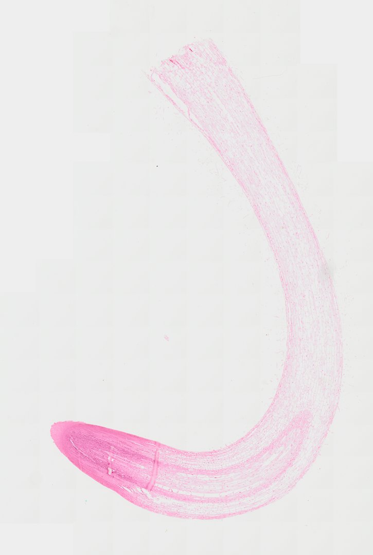
This is a lengthwise section through the tip of a corn root, stained with safranin. It shows the zones of a growing root, from the protective cap to the region where cells divide and stretch.
This is a lengthwise (longitudinal) section of a corn root tip (Zea mays), stained pink with safranin. At the very end is the root cap, a pad of cells that protects the tip as it pushes through soil. Just behind the cap is the meristem, a crowded zone of small, dividing cells with dark nuclei. Farther back, the cells get longer in the zone of elongation, and that stretching is what pushes the root deeper. Follow the root away from the tip to see how the cells change size and shape, and how the tissues sort into an outer layer, a cortex, and a central core that will carry water.
What to look for
- Root cap: A cap of cells covering the tip that protects the root as it pushes through soil.
- Meristem: Small, tightly packed cells just behind the cap, where cells divide.
- Dividing cells: Cells with dark-stained chromosomes, caught in mitosis, within the meristem.
- Zone of elongation: Farther back, cells become longer as they stretch.
- Developing tissues: Rows of cells forming the future epidermis, cortex, and central vascular tissue.
Questions and answers
What are the zones of a root tip?
A root tip has a root cap at the end, then a zone of cell division (the meristem), then a zone of elongation where cells stretch, and finally a zone of maturation where cells become specialized tissues.
What does the root cap do?
The root cap protects the delicate meristem as the root grows through the soil. Its outer cells wear off and are replaced, and it also helps the root sense gravity so it grows downward.
Compare it
- Category
- Botany & Plants
- Scan magnification
- 20x
- Stain
- Safranin
Viewing this slide in class
This is a scan of a real prepared slide, not a simulation or a drawing. On StudyScope you can pan from the whole slide down to individual structures, always in focus, and measure them in real micrometers with the calibrated ruler. Present it live and every student screen follows your view and annotations. Students join with a room code on Chromebooks, iPads, or any browser, with no accounts.