Capsella bursa-pastoris Flower Under the Microscope
Capsella bursa-pastoris Flower (Whole Mount), Capsella bursa-pastoris
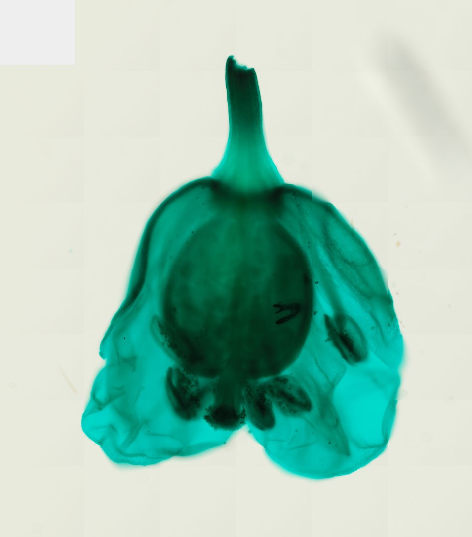
This is a whole flower of shepherd's purse (Capsella bursa-pastoris) mounted on a slide and stained green. Its small flower shows all four floral parts clearly, which makes it great for learning flower anatomy.
This whole mount shows an entire shepherd's purse (Capsella bursa-pastoris) flower, stained green. The flower stalk, or pedicel, is at the top of the view. The large, dark oval in the middle is the ovary, the base of the pistil where seeds form. Several anthers, the pollen sacs of the stamens, sit near the far end of the ovary, and some show dark pollen grains. Pale, see-through petals and sepals fan out around the edge, so students can see how all the parts connect.
What to look for
- Sepals: Small outer leaf-like parts at the base of the flower.
- Petals: Thin, delicate parts just inside the sepals.
- Stamens: Thin stalks (filaments) topped with anthers that hold pollen; in the mustard family there are usually six, four long and two short.
- Pistil and ovary: The central female part, with the ovary at its base.
- Pedicel: The small stalk holding the flower, with vascular strands running up into it.
Questions and answers
What are the parts of a flower under a microscope?
A flower has sepals on the outside, petals inside those, stamens (the male parts, made of filaments and anthers), and a pistil in the center (the female part, with an ovary at its base). A small flower like shepherd's purse shows all of these in one view.
Why is shepherd's purse used in botany classes?
Shepherd's purse is a small, common weed in the mustard family. Its flowers and developing seeds are classic teaching specimens because their structures are simple and easy to see on slides.
- Category
- Botany & Plants
- Scan magnification
- 20x
- Stain
- Fast Green
Viewing this slide in class
This is a scan of a real prepared slide, not a simulation or a drawing. On StudyScope you can pan from the whole slide down to individual structures, always in focus, and measure them in real micrometers with the calibrated ruler. Present it live and every student screen follows your view and annotations. Students join with a room code on Chromebooks, iPads, or any browser, with no accounts.