Corn Seed / Caryopsis Under the Microscope
Corn Seed / Caryopsis (Longitudinal Section), Zea mays
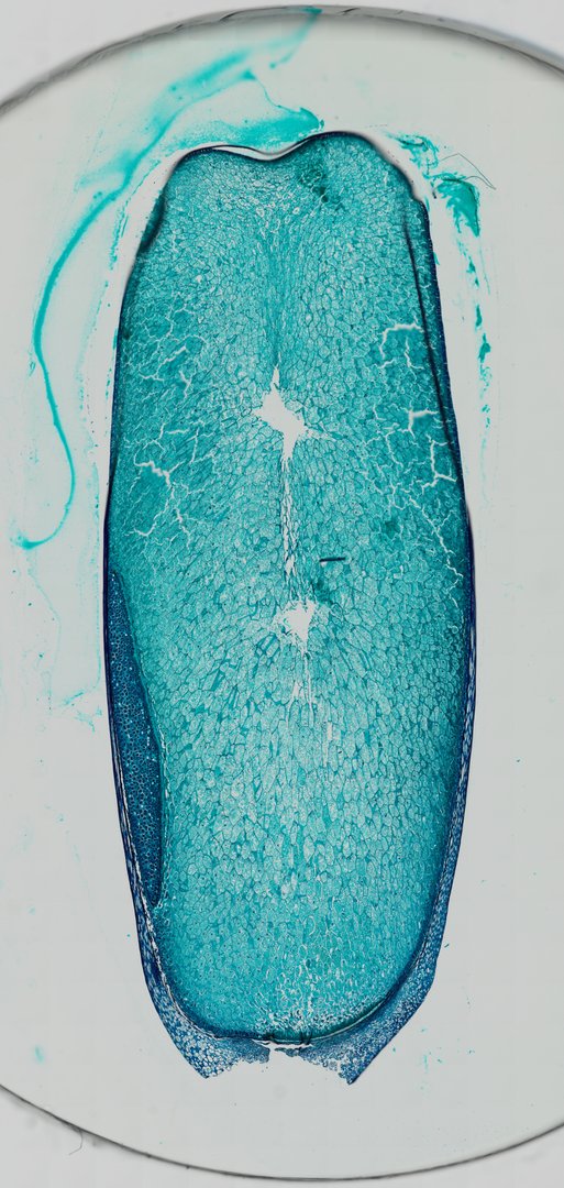
This is a lengthwise slice through a corn kernel stained with fast green. It shows the parts of a monocot seed, including the stored food and the tiny embryo plant.
This is a longitudinal section of a corn kernel (Zea mays), cut from top to bottom and stained with fast green. A corn kernel is a grain, a fruit with a single seed inside. The outer layer is the pericarp, a thin, tough wall that stains dark blue. Most of the kernel is endosperm, large cells packed with starch that feed the young plant when it sprouts. Along one lower side is part of the embryo, the scutellum, a shield-shaped seed leaf made of small, dense cells that takes up food from the endosperm.
What to look for
- Pericarp and seed coat: A thin outer layer wrapping the whole kernel.
- Endosperm: The large region filling most of the kernel, packed with stored starch.
- Aleurone layer: A single row of cells just inside the outer layer, at the edge of the endosperm.
- Scutellum: A shield-shaped structure (the single seed leaf) pressed against the endosperm.
- Coleoptile and plumule: The young shoot, wrapped in a protective sheath, pointing toward the top of the kernel.
- Radicle and coleorhiza: The young root, covered by its own sheath, pointing toward the base.
Questions and answers
What are the parts of a corn seed under a microscope?
A corn kernel has an outer pericarp and seed coat, a large starchy endosperm, and an embryo. The embryo includes the scutellum (seed leaf), a young shoot inside a sheath called the coleoptile, and a young root inside a sheath called the coleorhiza.
How is a corn seed different from a bean seed?
Corn is a monocot, so its embryo has one seed leaf (the scutellum) and most of its food is stored in the endosperm. A bean is a dicot, with two large seed leaves (cotyledons) that hold most of the stored food.
- Category
- Botany & Plants
- Scan magnification
- 20x
- Stain
- Fast Green
Viewing this slide in class
This is a scan of a real prepared slide, not a simulation or a drawing. On StudyScope you can pan from the whole slide down to individual structures, always in focus, and measure them in real micrometers with the calibrated ruler. Present it live and every student screen follows your view and annotations. Students join with a room code on Chromebooks, iPads, or any browser, with no accounts.