Corn Stem Cross Section Under the Microscope
Corn Stem (Cross Section), Zea mays
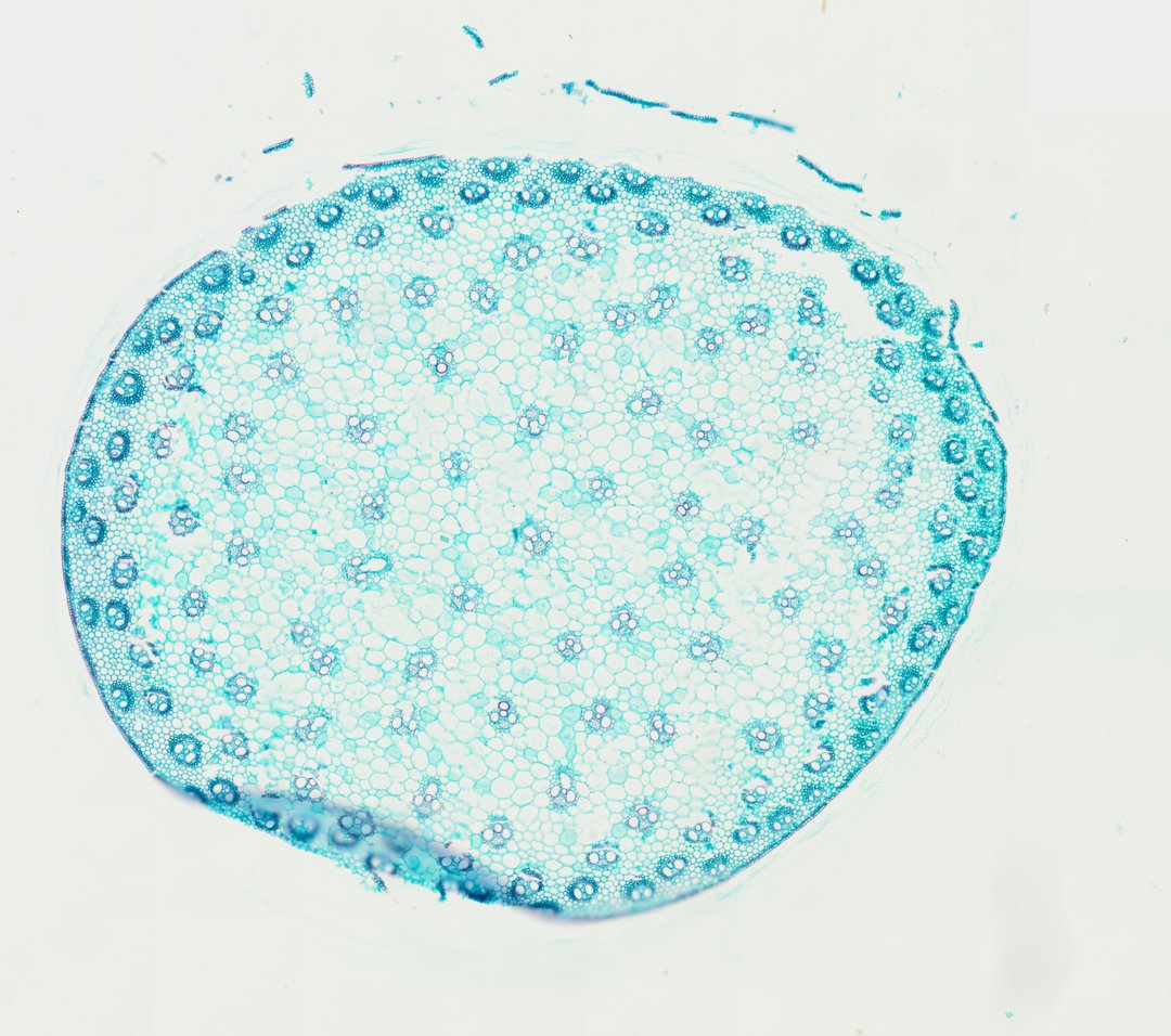
This is a cross section of a corn stem stained with fast green. It is the standard example of a monocot stem, where vascular bundles are scattered instead of arranged in a ring.
This is a cross section of a corn stem (Zea mays), stained with Fast Green. Corn is a monocot, so its vascular bundles are scattered all through the stem instead of forming one ring. The bundles are packed closer together near the outer edge and spread out toward the middle. Each bundle looks a bit like a face: two large xylem vessels (tubes that carry water) are the eyes, and the phloem, which carries sugar, sits between and above them. A ring of thick-walled cells wraps each bundle for support, and soft ground tissue fills the space between bundles.
Labeled corn stem cross section under the microscope
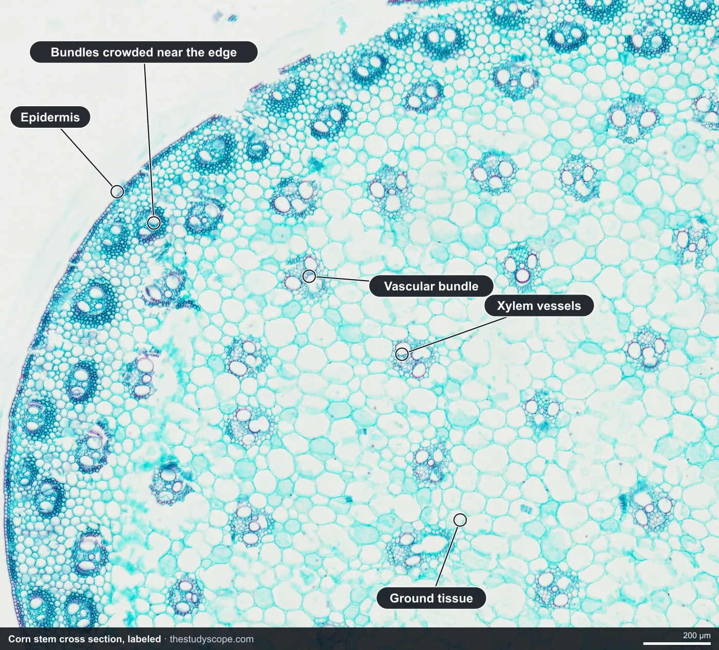
- Epidermis: Thin outer layer of small cells
- Bundles crowded near the edge: Vascular bundles packed close to the outside
- Vascular bundle: Scattered through the stem, not in a ring
- Xylem vessels: Large open tubes that carry water
- Ground tissue: Large thin-walled cells between bundles
What to look for
- Epidermis: A thin outer layer of small cells around the edge of the section.
- Scattered vascular bundles: Many small oval bundles spread through the stem, more crowded near the outer edge.
- Ground tissue: Large, thin-walled parenchyma cells filling the space between bundles.
- Xylem vessels: Two large, open vessels in each bundle, often with a smaller one between them, together looking a bit like a face.
- Phloem: A cluster of small cells on the outer side of each bundle.
- Bundle sheath: A ring of thick-walled cells surrounding each bundle.
Questions and answers
What does a monocot stem look like under a microscope?
In a monocot stem like corn, the vascular bundles are scattered all through the stem instead of forming one ring. There is no clear division into cortex and pith, and each bundle is wrapped in a sheath of thick-walled cells.
What is the difference between monocot and dicot stems?
Monocot stems have scattered vascular bundles and no ring of cambium, so they generally do not grow wider with wood. Dicot stems have bundles arranged in a ring, with pith in the center and cortex outside.
Why do corn vascular bundles look like a face?
Each bundle has two large xylem vessels that look like eyes, with smaller xylem cells and an air space below them and the phloem on the outer side. Students often describe it as a monkey face or skull, which makes it easy to recognize.
Compare it
- Category
- Botany & Plants
- Scan magnification
- 20x
- Stain
- Fast Green
Viewing this slide in class
This is a scan of a real prepared slide, not a simulation or a drawing. On StudyScope you can pan from the whole slide down to individual structures, always in focus, and measure them in real micrometers with the calibrated ruler. Present it live and every student screen follows your view and annotations. Students join with a room code on Chromebooks, iPads, or any browser, with no accounts.