Capsicum Peel Under the Microscope
Capsicum Peel (Whole Mount), Capsicum annuum
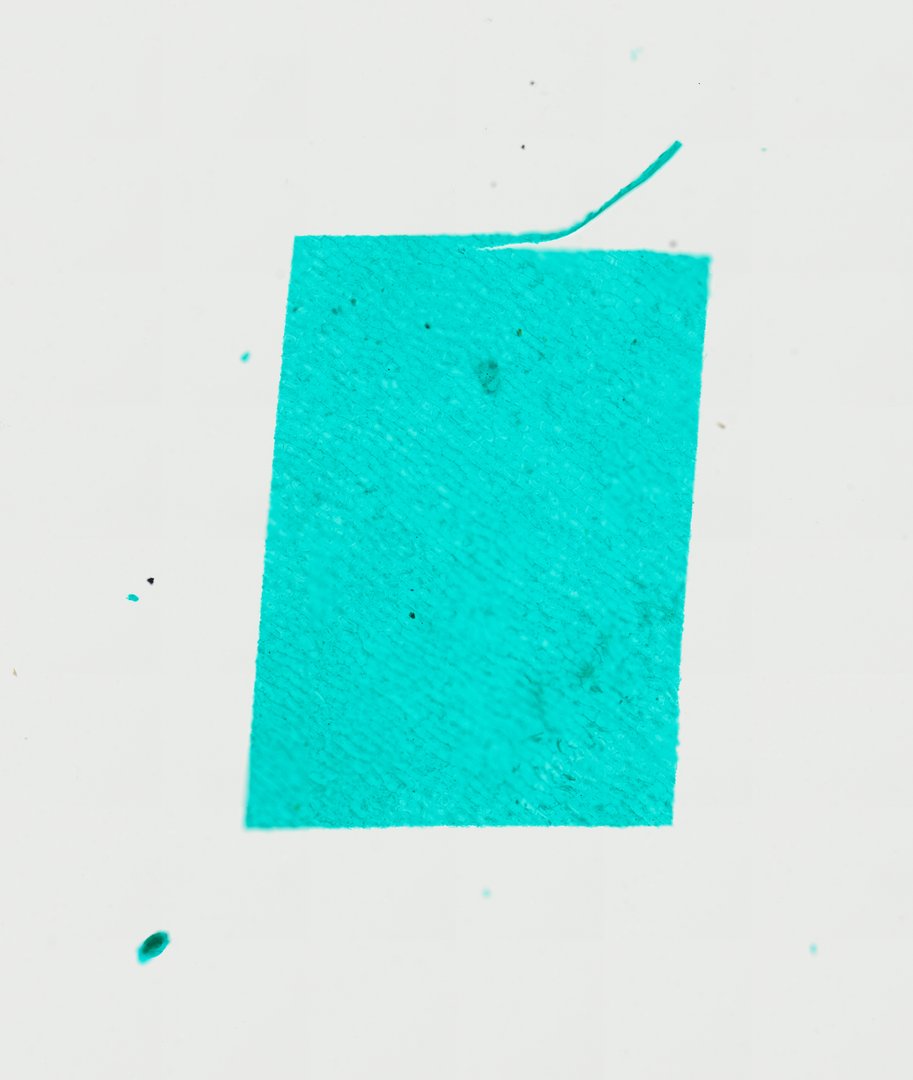
This is a thin peel of the skin of a pepper fruit, mounted flat and stained with fast green. It shows a neat sheet of plant epidermal cells with clear walls, a good contrast to leaf peels.
This is a whole mount of pepper skin, the thin outer layer peeled from a Capsicum annuum fruit and stained with fast green. It is one sheet of epidermis, the protective outer layer of cells. The cells are many-sided and fit tightly together like floor tiles, with no gaps between them. Their green cell walls make each outline easy to trace. Compare this peel with a leaf peel, which has many stomata, the small pores plants use for gas exchange.
What to look for
- Epidermal cells: Many-sided (polygonal) cells packed tightly like tiles, with no gaps between them.
- Cell walls: Clear green outlines around each cell, forming a mosaic pattern.
- Cell contents: Material inside some cells, which may include small bodies (plastids) and inclusions.
- Stomata: Occasional pairs of guard cells surrounding a small pore may appear in the sheet.
Questions and answers
What do pepper skin cells look like under a microscope?
Pepper skin (exocarp) cells are many-sided cells fitted tightly together like tiles. With fast green stain, their walls show up as clear green outlines.
How is a fruit peel different from a leaf peel?
Both are epidermis, a protective outer layer of cells. Leaf peels, especially from the underside, usually have many stomata for gas exchange, while fruit skin tends to have far fewer.
- Category
- Botany & Plants
- Scan magnification
- 20x
- Stain
- Fast Green
Viewing this slide in class
This is a scan of a real prepared slide, not a simulation or a drawing. On StudyScope you can pan from the whole slide down to individual structures, always in focus, and measure them in real micrometers with the calibrated ruler. Present it live and every student screen follows your view and annotations. Students join with a room code on Chromebooks, iPads, or any browser, with no accounts.