Amaranth Stem Under the Microscope (Longitudinal Section)
Amaranth Stem (Longitudinal Section), Amaranthus
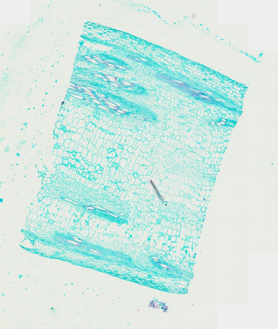
This is a thin lengthwise slice of an amaranth stem, a common herbaceous (non-woody) plant. Classes use it to see how plant tissues line up along a stem and how water-carrying xylem differs from the softer tissues around it.
This is a longitudinal section of an amaranth stem (Amaranthus), cut along its length and stained with safranin and fast green. Because of the cut, the cells look long and stretched instead of round. Most of the stem is ground tissue made of large, thin-walled parenchyma cells, with the epidermis, the outer skin layer, along each edge. Long vascular bundles run through it like stripes. Inside them, the xylem vessels, tubes that carry water up from the roots, show spiral and ring-shaped wall thickenings stained pink to purple, while the other cell walls stain green.
What to look for
- Epidermis: A thin outer layer of small cells running along each edge of the section.
- Parenchyma (ground tissue): Long, thin-walled cells filling most of the section, stained green by fast green.
- Xylem vessels: Long tubes stained red by safranin, often with spiral or ring-shaped thickenings in their walls.
- Phloem: Narrow, green-stained cells running right alongside the xylem strands.
- Vascular bundles: Stripes of xylem and phloem running parallel to the length of the stem.
Questions and answers
What does a stem longitudinal section look like under a microscope?
A longitudinal section is cut along the length of the stem, so cells look long and stretched instead of round. Vascular bundles show up as parallel stripes, and xylem vessels look like long tubes with spiral or ring-shaped wall patterns.
Why is xylem red and other tissue green on this slide?
The slide is stained with safranin and fast green. Safranin binds to lignin, the stiff material in xylem walls, and turns it red, while fast green colors the thinner cellulose walls of other cells green.
What is the difference between a longitudinal section and a cross section?
A cross section is cut straight across the stem, like slicing a carrot into coins, so you see tissues as circles and rings. A longitudinal section is cut lengthwise, so you see the same tissues as long strips.
- Category
- Botany & Plants
- Scan magnification
- 20x
- Stain
- Safranin / Fast Green
Viewing this slide in class
This is a scan of a real prepared slide, not a simulation or a drawing. On StudyScope you can pan from the whole slide down to individual structures, always in focus, and measure them in real micrometers with the calibrated ruler. Present it live and every student screen follows your view and annotations. Students join with a room code on Chromebooks, iPads, or any browser, with no accounts.