Young Root of Broad Bean Under the Microscope
Young Root of Broad Bean (Cross Section), Vicia faba
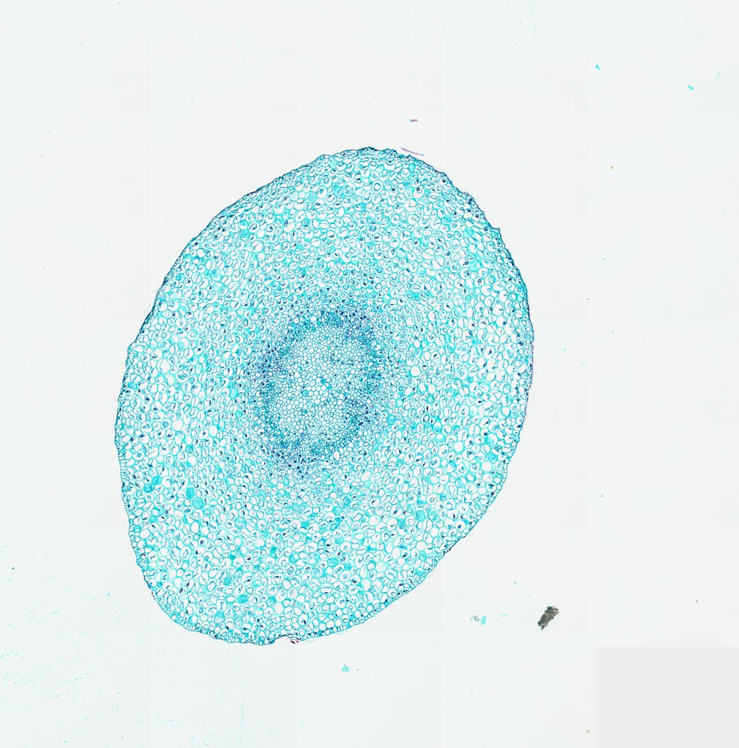
This is a cross section of a young broad bean root stained with fast green. It shows the classic tissue layers of a young dicot root.
This is a cross section of a young broad bean (Vicia faba) root, stained with fast green. The outer edge is the epidermis, a single layer of cells that takes in water from the soil. Most of the root is cortex, a wide zone of large, round cells that store food. At the center is the stele, the root's core, wrapped in a ring of small cells called the endodermis, which controls what water and minerals get inside. Within the stele, groups of xylem (tubes that carry water up) sit between groups of phloem (tubes that carry sugar), a pattern students can compare with a stem.
What to look for
- Epidermis: The outer layer of cells, which may have root hairs.
- Cortex: A wide zone of large, round cells with small air spaces between them.
- Endodermis: A single ring of cells at the inner edge of the cortex.
- Pericycle: A thin layer just inside the endodermis.
- Xylem and phloem: Groups of xylem and phloem that alternate around the center of the root.
- Pith: Small central area of thin-walled cells, in the middle of the vascular tissue.
Questions and answers
What are the parts of a root cross section?
From the outside in, a root has an epidermis, a wide cortex, a ring of endodermis, a pericycle, and a central core of xylem and phloem. In a young dicot root, xylem and phloem alternate around the center.
Where do side roots come from?
Side roots start in the pericycle, the layer just inside the endodermis. They grow outward through the cortex and break through the epidermis.
- Category
- Botany & Plants
- Scan magnification
- 20x
- Stain
- Fast Green
Viewing this slide in class
This is a scan of a real prepared slide, not a simulation or a drawing. On StudyScope you can pan from the whole slide down to individual structures, always in focus, and measure them in real micrometers with the calibrated ruler. Present it live and every student screen follows your view and annotations. Students join with a room code on Chromebooks, iPads, or any browser, with no accounts.