Tilia Stem Cross Section Under the Microscope
Tilia Stem (Cross Section), Tilia
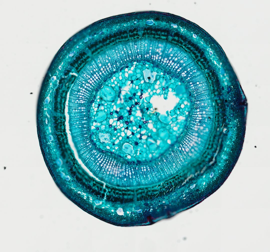
This is a cross section of a basswood (Tilia) stem stained with fast green. It is the classic slide for woody stem growth, showing annual rings of wood and the bark layers around them.
This is a cross section of a young basswood stem (Tilia), stained with fast green. At the center is the pith, a core of large, soft cells. Around it is a thick ring of wood, or secondary xylem, made of rows of small cells that carry water. Thin lines called rays cross the wood like spokes on a wheel. Outside the wood is a darker band of phloem, which carries food, and on the very outside are the cortex and cork that protect the stem. The layers show how a woody stem grows wider each year.
What to look for
- Pith: Soft tissue in the very center of the stem.
- Secondary xylem (wood): A wide zone of packed cells around the pith, often showing annual rings.
- Annual rings: Rings in the wood, each made of larger spring cells and smaller summer cells.
- Rays: Thin lines of cells running outward from the center, like spokes.
- Vascular cambium: A thin ring of small cells between the wood and the phloem.
- Secondary phloem: Triangle-shaped bands of phloem between rays that widen outward, a well-known basswood feature.
Questions and answers
How do you count tree rings on a Tilia stem slide?
Each annual ring has a band of larger cells made in spring and smaller cells made in late summer. Count the lines where small cells meet big ones, from the pith outward, to find how many years of growth the stem has.
What are the parts of a woody stem cross section?
From the center outward, a woody stem has pith, wood (secondary xylem) with annual rings and rays, the vascular cambium, the secondary phloem, and the cortex and cork on the outside.
Why is Tilia used for woody stem slides?
Basswood stems show annual rings, rays, and phloem clearly in a young stem. The triangle-shaped phloem bands make it especially easy to find the phloem and cambium.
Compare it
- Category
- Botany & Plants
- Scan magnification
- 20x
- Stain
- Fast Green
Viewing this slide in class
This is a scan of a real prepared slide, not a simulation or a drawing. On StudyScope you can pan from the whole slide down to individual structures, always in focus, and measure them in real micrometers with the calibrated ruler. Present it live and every student screen follows your view and annotations. Students join with a room code on Chromebooks, iPads, or any browser, with no accounts.