Spirogyra Under the Microscope
Spirogyra (Whole Mount), Spirogyra
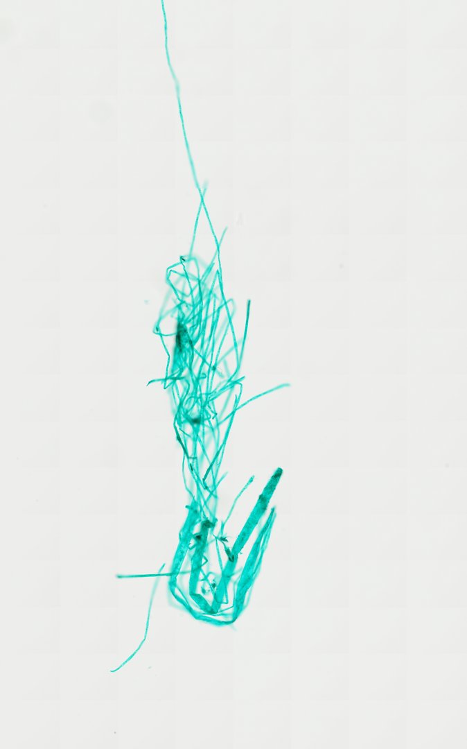
This is Spirogyra, a thread-like green alga, mounted whole and stained with fast green. Its spiral chloroplasts are among the most recognizable structures in biology class.
This whole mount shows Spirogyra, a green alga that grows as long, unbranched threads called filaments. Each filament is a chain of cylinder-shaped cells joined end to end, with thin cross walls between them. A dark spot near the middle of many cells is the nucleus. Spirogyra is named for its ribbon-shaped chloroplast, which winds in a spiral inside each cell and does the work of photosynthesis. The Fast Green stain colors the filaments blue-green, making it easy to follow single threads through the tangle.
Labeled spirogyra under the microscope
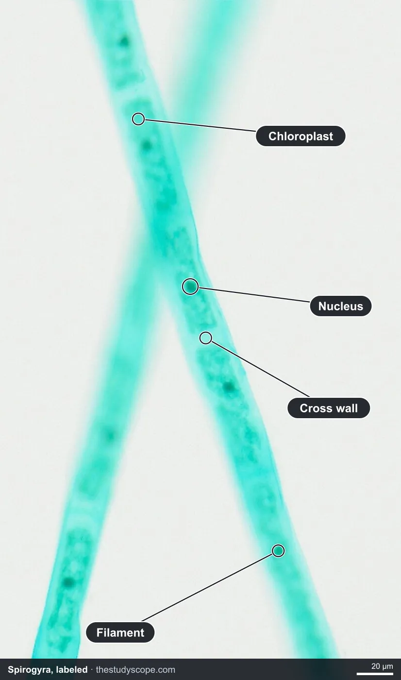
- Chloroplast: Ribbon that makes food by photosynthesis
- Nucleus: Dark spot held near the middle of the cell
- Cross wall: Where one cell ends and the next begins
- Filament: Chain of cells joined end to end
What to look for
- Filaments: Long, unbranched threads made of cells joined end to end.
- Cells: Cylinder-shaped cells, with walls marking where one ends and the next begins.
- Spiral chloroplasts: Ribbon-shaped chloroplasts that twist in a spiral along the inside of each cell.
- Pyrenoids: Small round bodies spaced along the chloroplast ribbon, where starch is made.
- Nucleus: A nucleus near the middle of each cell, held in place by thin strands of cytoplasm.
Questions and answers
What does Spirogyra look like under a microscope?
Spirogyra looks like long green threads made of cylinder-shaped cells. Inside each cell is a ribbon-shaped chloroplast that winds in a spiral, dotted with small round pyrenoids.
How does Spirogyra reproduce?
Spirogyra can grow by breaking into pieces that keep growing. It can also reproduce sexually by conjugation, when two filaments line up side by side, form bridges between cells, and the contents of one cell move into the other.
Compare it
- Category
- Botany & Plants
- Scan magnification
- 20x
- Stain
- Fast Green
Viewing this slide in class
This is a scan of a real prepared slide, not a simulation or a drawing. On StudyScope you can pan from the whole slide down to individual structures, always in focus, and measure them in real micrometers with the calibrated ruler. Present it live and every student screen follows your view and annotations. Students join with a room code on Chromebooks, iPads, or any browser, with no accounts.