Pine Stem Cross Section Under the Microscope
Pine Stem (Cross Section), Pinus
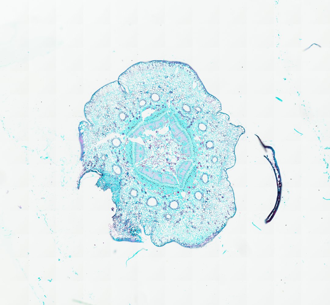
This is a cross section of a young pine stem stained with safranin and fast green. It shows how conifer stems are built, with wood made of tracheids and resin ducts running through the tissue.
This is a cross section of a young pine stem (Pinus), stained with safranin and fast green. The center is the pith, made of large, thin-walled cells, and around it sits a ring of wedge-shaped xylem, the wood that carries water, made of small cells in neat rows. Just outside the xylem are the cambium, a thin layer that makes new wood, and the phloem, which carries sugars. The outer cortex holds many round resin ducts, each ringed by small cells that release sticky resin to seal wounds. Pine has no large vessels in its wood, which sets it apart from flowering plants.
What to look for
- Pith: Large, thin-walled cells in the very center of the stem.
- Xylem: A ring of red-stained, thick-walled cells (tracheids) around the pith.
- Vascular cambium: A thin layer of small cells between the xylem and the phloem.
- Phloem: A green-stained band just outside the cambium.
- Resin ducts: Round canals lined with small cells, found in the cortex and sometimes in the xylem.
- Cortex and outer layers: Cells between the phloem and the surface, covered by epidermis or early bark.
Questions and answers
What does a pine stem look like under a microscope?
A young pine stem has pith in the center, a ring of xylem (wood) stained red, a thin cambium layer, phloem, and a cortex with round resin ducts. The xylem is made mostly of tracheids arranged in neat rows.
How is pine wood different from oak or maple wood?
Pine is a softwood, so its wood is made almost entirely of tracheids and has no vessels. Hardwoods like oak and maple have large water-carrying vessels that show up as bigger holes in a cross section.
- Category
- Botany & Plants
- Scan magnification
- 20x
- Stain
- Safranin / Fast Green
Viewing this slide in class
This is a scan of a real prepared slide, not a simulation or a drawing. On StudyScope you can pan from the whole slide down to individual structures, always in focus, and measure them in real micrometers with the calibrated ruler. Present it live and every student screen follows your view and annotations. Students join with a room code on Chromebooks, iPads, or any browser, with no accounts.