Pine Needle Cross Section Under the Microscope
Pine Needle (Cross Section), Pinus
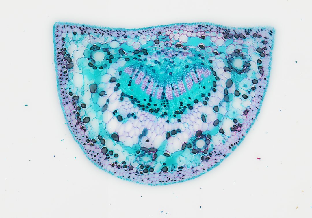
This is a cross section of a pine needle stained with safranin and fast green. Pine needles are leaves built to save water in cold or dry conditions, and this slide shows many of those adaptations.
This is a cross section of a pine needle (Pinus), stained with safranin and fast green. The needle is covered by a thick, waxy cuticle and a layer of thick-walled cells that help it keep water in. Under that, the mesophyll cells carry out photosynthesis, and several round resin ducts, rings of cells around an open center, carry sticky resin that helps protect the tree. In the middle, a ring of cells called the endodermis surrounds the vascular tissue, where xylem carries water and phloem carries food. These features help pines survive dry and freezing weather.
What to look for
- Cuticle and epidermis: A thick waxy coat over a layer of thick-walled cells.
- Hypodermis: Thick-walled, often red-stained cells just under the epidermis that add strength.
- Sunken stomata: Stomata set down into the surface, which reduces water loss.
- Mesophyll: Photosynthetic cells with folded walls that increase surface area.
- Resin ducts: Round canals lined with small cells, spaced through the mesophyll.
- Endodermis and vascular tissue: A ring of cells around the central vascular region, with xylem stained red.
Questions and answers
What are the parts of a pine needle cross section?
A pine needle has a thick cuticle, epidermis, and hypodermis on the outside, sunken stomata, mesophyll cells with folded walls, resin ducts, and a central vascular region surrounded by an endodermis.
How are pine needles adapted to save water?
Pine needles have a small surface area, a thick waxy cuticle, a tough layer of thick-walled cells under the skin, and stomata sunk below the surface. Together, these reduce water loss in dry or freezing weather.
Compare it
- Category
- Botany & Plants
- Scan magnification
- 20x
- Stain
- Safranin and Fast Green
Viewing this slide in class
This is a scan of a real prepared slide, not a simulation or a drawing. On StudyScope you can pan from the whole slide down to individual structures, always in focus, and measure them in real micrometers with the calibrated ruler. Present it live and every student screen follows your view and annotations. Students join with a room code on Chromebooks, iPads, or any browser, with no accounts.