Lower Epidermis of Broad Bean Leaf Under the Microscope
Lower Epidermis of Broad Bean Leaf (Whole Mount), Vicia faba
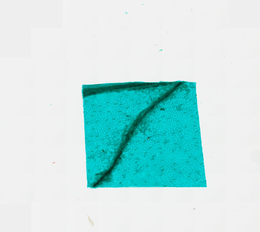
This is a peel of the lower surface of a broad bean leaf, mounted flat and stained with fast green. It is one of the best slides for seeing stomata and the guard cells that open and close them.
This whole mount shows the lower epidermis, the thin skin on the underside of a broad bean (Vicia faba) leaf, stained with Fast Green. Most of the sheet is made of pavement cells with wavy walls that fit together like puzzle pieces. Scattered among them are many stomata, small pores that let carbon dioxide in and water vapor out. Each pore sits between two bean-shaped guard cells that swell or shrink to open and close it. Dark strands of a leaf vein cross the peel, and students can count the stomata in one area and compare the count with other leaves.
Labeled broad bean leaf epidermis under the microscope
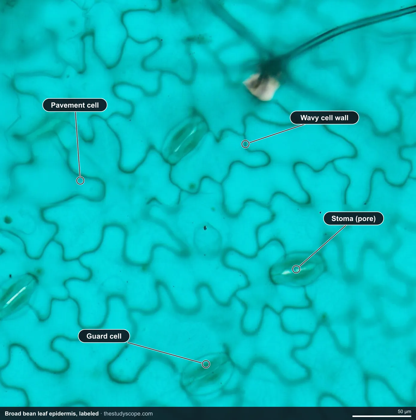
- Stoma (pore): Opening that lets gases in and out
- Guard cell: Bean-shaped cell that opens and closes the pore
- Pavement cell: Wavy-walled cell that fits like a puzzle piece
- Wavy cell wall: Walls fit together like puzzle pieces
What to look for
- Pavement cells: Epidermal cells with wavy walls that fit together like puzzle pieces.
- Stomata: Small pores scattered across the surface.
- Guard cells: Pairs of kidney-shaped cells around each pore.
- Veins: Long, narrow cells marking where veins run under the epidermis.
Questions and answers
What do stomata look like under a microscope?
A stoma looks like a small slit or pore between two bean-shaped guard cells. On a leaf peel, they are scattered among the puzzle-piece shaped epidermal cells.
Why are there more stomata on the bottom of a leaf?
The underside of a leaf is shaded and cooler, so less water is lost through stomata there. Many land plants have most of their stomata on the lower surface for this reason.
How do guard cells open and close a stoma?
When guard cells take in water, they swell and bow apart, opening the pore. When they lose water, they go limp and the pore closes, which helps the plant save water.
Compare it
- Category
- Botany & Plants
- Scan magnification
- 20x
- Stain
- Fast Green
Viewing this slide in class
This is a scan of a real prepared slide, not a simulation or a drawing. On StudyScope you can pan from the whole slide down to individual structures, always in focus, and measure them in real micrometers with the calibrated ruler. Present it live and every student screen follows your view and annotations. Students join with a room code on Chromebooks, iPads, or any browser, with no accounts.