Human Blood Smear Under the Microscope
Human Blood Smear, Homo sapiens
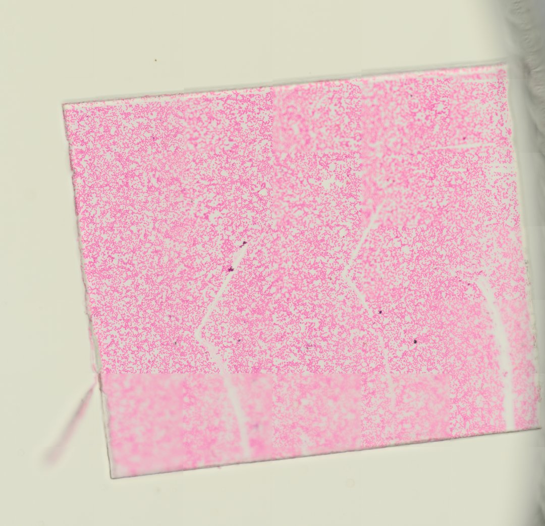
This is a thin smear of human blood stained with Wright-Giemsa. It is the standard slide for identifying red blood cells, the different types of white blood cells, and platelets.
This is a smear of human blood (Homo sapiens), spread thin on a slide and stained with Wright-Giemsa. Most of the field is filled with red blood cells, small pink disks with pale centers and no nucleus. They carry oxygen to the body. Here and there, larger cells with dark purple nuclei stand out, and these are white blood cells, which help fight infection. Comparing how many red cells there are to how few white cells gives a sense of the balance in healthy blood.
Labeled human blood smear under the microscope
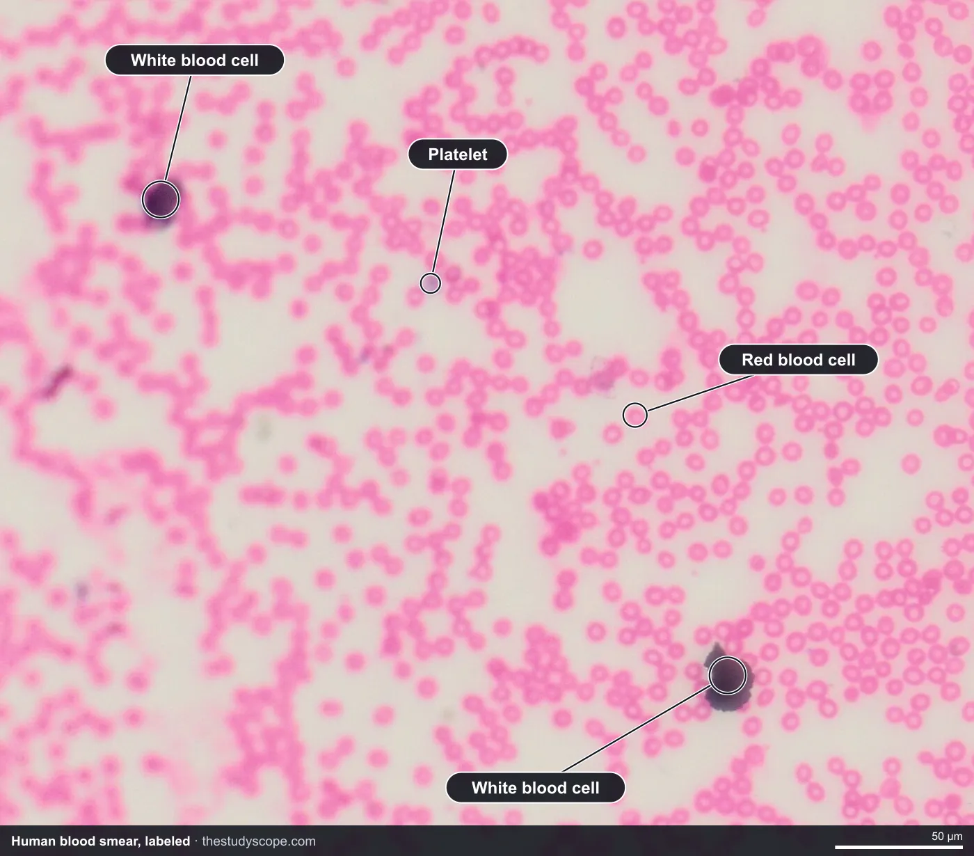
- White blood cell: Larger cell with a dark purple nucleus
- Platelet: Tiny purple fragment that helps blood clot
- Red blood cell: Pink disk with a pale center and no nucleus
What to look for
- Red blood cells: Round, pink cells with no nucleus and a paler center, filling most of the slide.
- Neutrophils: The most common white blood cell, with a purple nucleus split into several lobes.
- Lymphocytes: Small white blood cells with a large, round, dark nucleus and a thin rim of blue cytoplasm.
- Monocytes: The largest white blood cells, with a kidney- or bean-shaped nucleus.
- Eosinophils and basophils: Less common white blood cells with orange-red granules (eosinophils) or dark purple granules (basophils).
- Platelets: Tiny purple fragments scattered between the red blood cells.
Questions and answers
What does human blood look like under a microscope?
Human blood is mostly small, round, pink red blood cells with pale centers and no nucleus. Scattered among them are larger white blood cells with purple nuclei and tiny purple platelets.
How do you identify white blood cells on a blood smear?
Look at the nucleus and granules. Neutrophils have a lobed nucleus, lymphocytes have a big round nucleus, monocytes have a bean-shaped nucleus, eosinophils have orange-red granules, and basophils have dark purple granules.
How big are human red blood cells?
Human red blood cells are about 7 to 8 micrometers across. Because their size is so consistent, they are handy for estimating the size of other cells on a slide.
Compare it
- Category
- Vertebrates
- Scan magnification
- 20x
- Stain
- Wright-Giemsa
Viewing this slide in class
This is a scan of a real prepared slide, not a simulation or a drawing. On StudyScope you can pan from the whole slide down to individual structures, always in focus, and measure them in real micrometers with the calibrated ruler. Present it live and every student screen follows your view and annotations. Students join with a room code on Chromebooks, iPads, or any browser, with no accounts.