Three Types of Bacteria Smear Under the Microscope
Three Types of Bacteria Smear, Bacteria
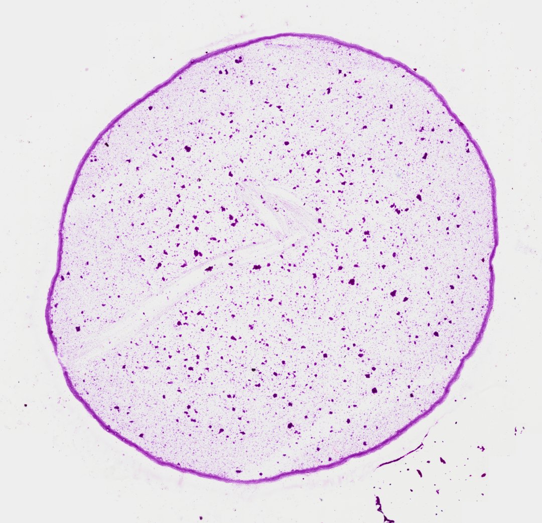
This is a smear of three kinds of bacteria stained purple with crystal violet. It shows the three basic bacterial shapes: round cocci, rod-shaped bacilli, and spiral spirilla.
This is a bacteria smear stained with crystal violet, a purple dye that makes tiny cells easy to see. The slide is meant to show the three basic shapes of bacteria: cocci (round), bacilli (rod-shaped), and spirilla (spiral). In the parts of this smear shown here, most of the cells are short rods, spread out alone or packed into dark clumps. Each rod is only a few micrometers long. Shape is one of the first clues scientists use to sort and identify bacteria.
Labeled bacteria smear under the microscope
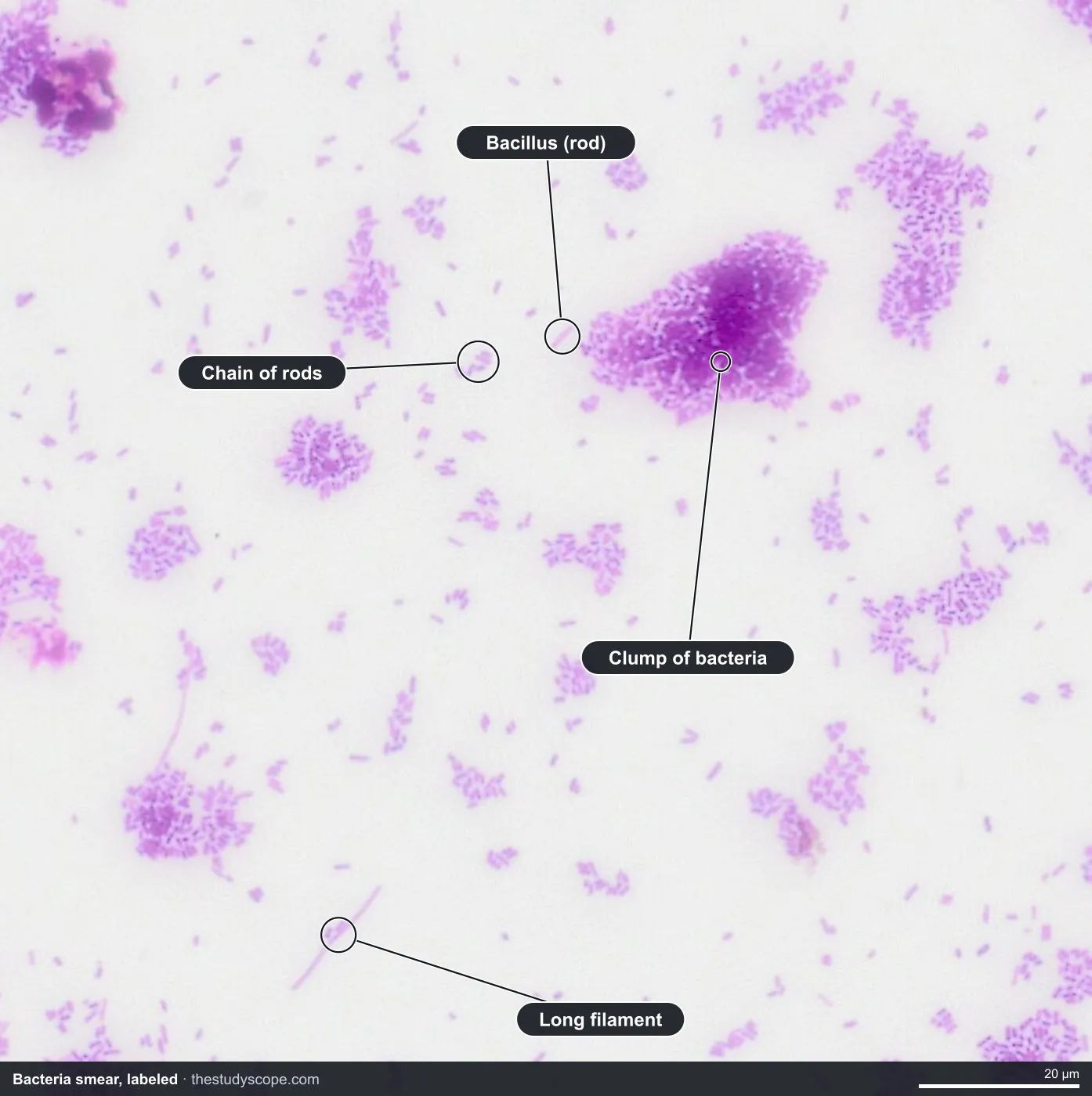
- Bacillus (rod): A single rod-shaped cell
- Chain of rods: Rods stuck end to end or side by side
- Clump of bacteria: Many rods packed together
- Long filament: A long, thin thread of cells
What to look for
- Cocci: Tiny round cells, alone, in pairs, in chains, or in clusters.
- Bacilli: Short rod-shaped cells.
- Spirilla: Longer cells with a wavy or corkscrew shape.
Questions and answers
What are the three shapes of bacteria?
The three basic shapes are cocci (round), bacilli (rod-shaped), and spirilla (spiral). Shape is one of the first clues microbiologists use to identify bacteria.
Why do you need high magnification to see bacteria?
Most bacteria are only about 1 to 5 micrometers long, much smaller than human cells. At low power they look like dust, so you need the highest magnification to see their shapes.
Is this slide a Gram stain?
No. This slide uses one stain, crystal violet, so all the bacteria look purple. A Gram stain uses several steps to sort bacteria into purple (Gram-positive) and pink (Gram-negative) groups.
Compare it
- Category
- Microbiology
- Scan magnification
- 40x
- Stain
- Crystal Violet
Viewing this slide in class
This is a scan of a real prepared slide, not a simulation or a drawing. On StudyScope you can pan from the whole slide down to individual structures, always in focus, and measure them in real micrometers with the calibrated ruler. Present it live and every student screen follows your view and annotations. Students join with a room code on Chromebooks, iPads, or any browser, with no accounts.