Paramecium Under the Microscope
Paramecium (Whole Mount), Paramecium
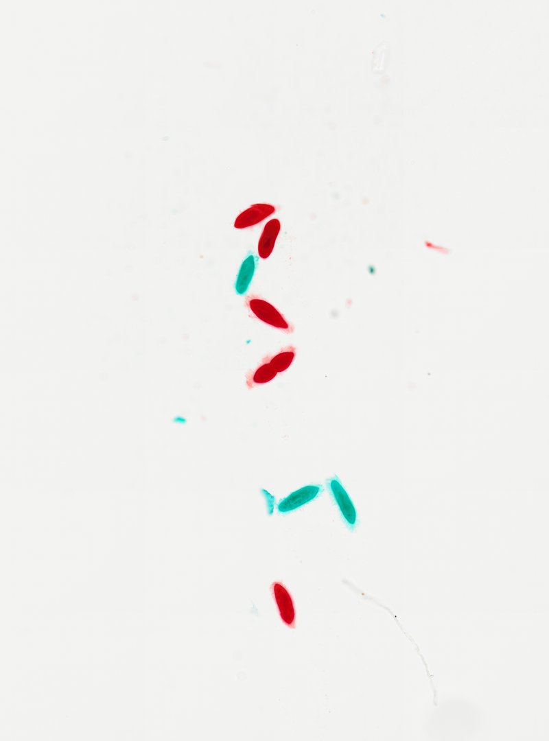
This slide shows many Paramecium cells mounted whole and stained. Paramecium is a single-celled organism covered in cilia and is one of the most familiar protists in biology class.
This whole mount shows several Paramecium, single-celled protists that live in fresh water. Each cell has a slipper shape, rounded at one end and slightly narrower at the other. Some cells are stained red and others teal, which makes their outlines easy to see. Look closely at the edges to find cilia, tiny hairs that cover the cell and beat like oars to move it through water. Some cells also show small round food vacuoles inside, where the Paramecium digests its food.
Labeled paramecium under the microscope
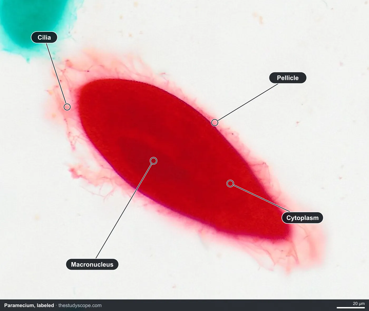
- Cilia: Tiny hairs that beat like oars
- Macronucleus: Large, darker nucleus near the middle
- Pellicle: Firm outer covering of the cell
- Cytoplasm: Material filling the cell
What to look for
- Slipper shape: An oval cell with one end more rounded and the other more tapered.
- Oral groove: A dent along one side of the cell where food is swept in.
- Macronucleus: A large, dark-staining nucleus near the middle of the cell.
- Micronucleus: A much smaller nucleus next to the macronucleus, which can be hard to see.
- Food vacuoles: Small round bubbles inside the cell holding food being digested.
- Cilia: Very fine hairs covering the surface, often hard to see on stained slides.
Questions and answers
What are the parts of a Paramecium?
A Paramecium has cilia covering its surface, an oral groove for taking in food, food vacuoles, contractile vacuoles that pump out extra water, and two kinds of nuclei: a large macronucleus and a small micronucleus.
How big is a Paramecium?
Many common Paramecium species are around 0.2 millimeters long, so they are just barely visible to the naked eye as tiny specks in pond water.
How does a Paramecium move?
Paramecium swims by beating thousands of tiny cilia in waves. The cilia also sweep food into the oral groove.
Compare it
- Category
- Microbiology
- Scan magnification
- 20x
- Stain
- Various
Viewing this slide in class
This is a scan of a real prepared slide, not a simulation or a drawing. On StudyScope you can pan from the whole slide down to individual structures, always in focus, and measure them in real micrometers with the calibrated ruler. Present it live and every student screen follows your view and annotations. Students join with a room code on Chromebooks, iPads, or any browser, with no accounts.