Honeybee Wing Under the Microscope
Honeybee Wing (Whole Mount), Apis mellifera
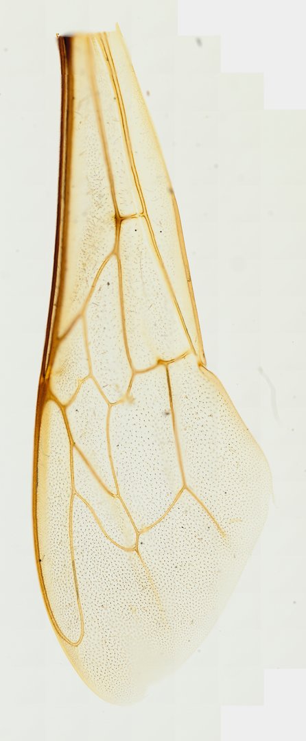
This slide shows honeybee wings mounted whole and unstained. The vein pattern is used to identify bees, and the wings show how insects with two pairs of wings can hook them together in flight.
This is a whole mount of a honeybee wing (Apis mellifera), shown unstained in its natural color. The wing is a thin, clear membrane held stiff by a network of brown, hardened veins. The veins join to form closed shapes called cells, and their pattern is used to identify bees and other insects. The thick, dark vein along the front edge makes that edge strong for flight, and tiny hairs cover the membrane as fine dots. Students can trace the veins and count the cells, then compare this wing with a fly or butterfly wing.
What to look for
- Wing membrane: The clear sheet of the wing.
- Veins: Brown, hardened lines that support the membrane.
- Cells: Closed areas of membrane surrounded by veins, in a pattern used to identify bee species.
- Hamuli: A row of tiny hooks on the front edge of the hind wing that lock it to the forewing, if the hind wing is included.
- Microtrichia: Tiny hairs covering the membrane, visible at high power.
Questions and answers
How do honeybee wings work together?
Honeybees have two pairs of wings. A row of tiny hooks called hamuli on the front edge of each hind wing catches a fold on the forewing, so both wings on each side flap as one surface.
What does a bee wing look like under a microscope?
A bee wing is a clear membrane with a pattern of brown veins that form closed cells. At higher magnification, you can see tiny hairs covering the whole surface.
- Category
- Invertebrates
- Scan magnification
- 20x
- Stain
- Unstained
Viewing this slide in class
This is a scan of a real prepared slide, not a simulation or a drawing. On StudyScope you can pan from the whole slide down to individual structures, always in focus, and measure them in real micrometers with the calibrated ruler. Present it live and every student screen follows your view and annotations. Students join with a room code on Chromebooks, iPads, or any browser, with no accounts.