Cotton Root (Cross Section)
Gossypium hirsutum
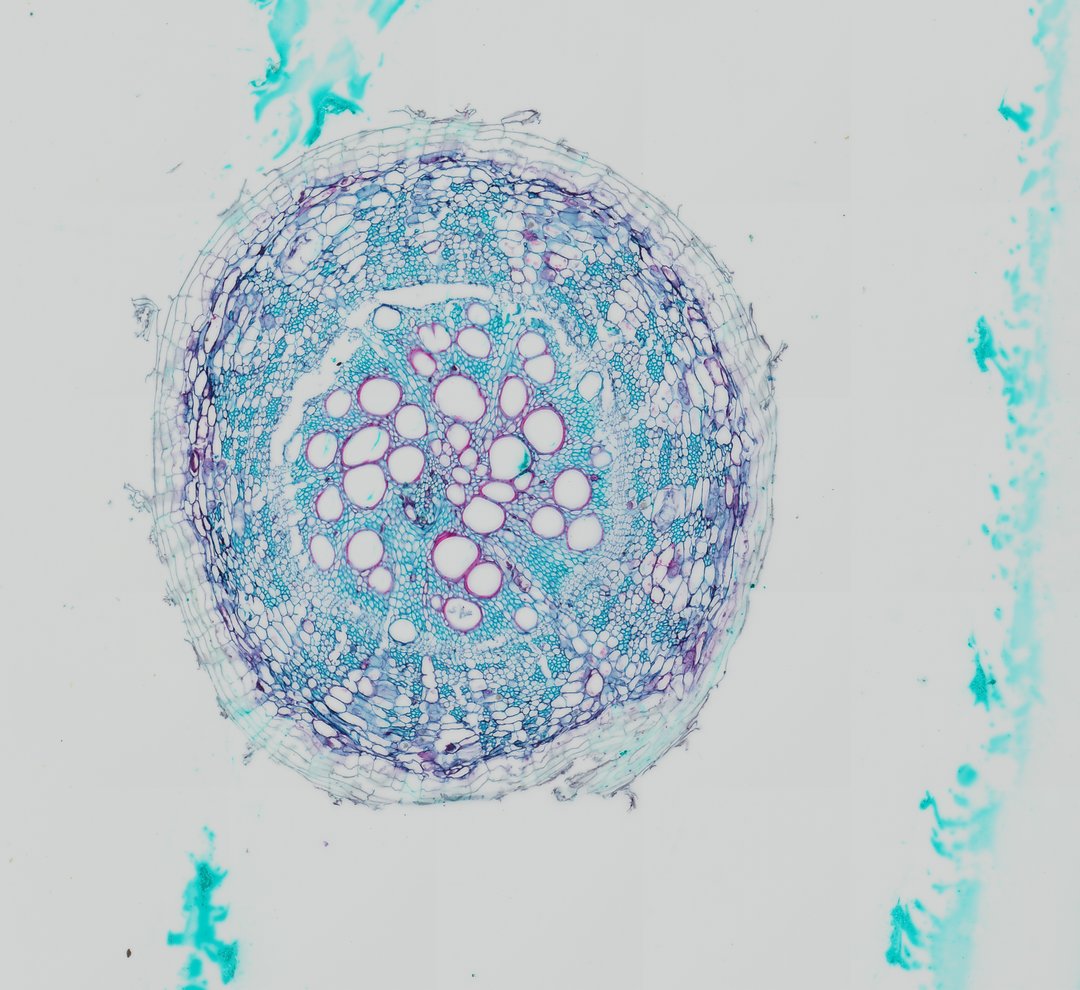
This cross section of a cotton root (Gossypium hirsutum c.s.) reveals the fundamental anatomical organization of a young dicotyledonous root undergoing primary growth and early vascular development. The outermost boundary is composed of an epidermis and underlying cortical parenchyma cells that store reserves and facilitate lateral water transport. Centrally located within the vascular cylinder (stele), a prominent arrangement of xylem vessels displays thick, lignified secondary cell walls stained red/magenta by safranin, interspaced with primary and secondary phloem elements stained blue-green. Surrounding the vascular tissue is the endodermis, which features Casparian strips regulating apoplastic transport into the vascular core. The distinct star-shaped or tetrarch/polyarch vascular arrangement and peripheral pericycle layer highlight structural features characteristic of Malvaceae root anatomy.
- Category
- Botany & Plants
- Magnification
- 20x
- Stain
- Safranin and Fast Green
- Tags
- cotton root cross section, Gossypium hirsutum, dicot root, xylem vessels, phloem, stele, endodermis, Casparian strip, pericycle, root cortex, parenchyma, lignified cell wall, plant tissue cross section, vascular bundle, plant histology, botanical microtechnique, safranin fast green, brightfield microscopy, Malvaceae
Explore this slide with deep zoom, save it to your library, and present it live to students on StudyScope. Create a teacher account or browse the full catalog.