Oyster Gill (Cross Section)
Crassostrea virginica
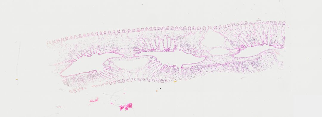
A cross section (c.s.) of an oyster gill (labeled Oyster gill c.s.). The slide demonstrates the complex bivalve demibranch architecture, showing w-shaped ciliated gill filaments, ostia, and internal water tubes involved in both respiration and filter feeding.
- Category
- Invertebrates
- Magnification
- 20x
- Stain
- H&E
- Tags
- Oyster gill c.s., Oyster gill cross section, Oyster gill t.s., Oyster gill transverse section, Oyster gill, Crassostrea virginica, Crassostrea, Oyster, Bivalvia, Mollusca, Gill filament, Ciliated epithelium, Demibranch, Water tubes, Respiration, Filter feeding, Invertebrate histology, Cross section, H&E stain
Explore this slide with deep zoom, save it to your library, and present it live to students on StudyScope. Create a teacher account or browse the full catalog.