Small Intestine (Cross Section)
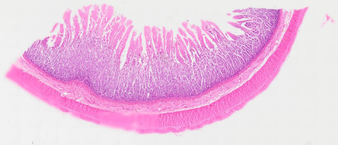
This cross-sectional histological preparation displays the characteristic four-layered wall architecture of the small intestine stained with Hematoxylin and Eosin (H&E). Extending into the lumen are tall, finger-like villi lined by simple columnar epithelium consisting primarily of absorptive enterocytes with apical microvilli (striated border) and interspersed mucin-secreting goblet cells. At the base of the villi, tubular invaginations form the crypts of Lieberkühn within the lamina propria. Beneath the mucosa, the thin muscularis mucosae separates the inner layer from the connective tissue submucosa. Surrounding the submucosa is a thick muscularis externa composed of inner circular and outer longitudinal smooth muscle layers, bounded externally by the visceral peritoneum (serosa).
- Category
- Histology and Tissue
- Magnification
- 20x
- Stain
- H&E
- Tags
- small intestine cross section, small intestine, intestinal villi, crypts of Lieberkühn, simple columnar epithelium, enterocytes, goblet cells, lamina propria, muscularis mucosae, submucosa, muscularis externa, smooth muscle, digestive tract histology, gastrointestinal tract, H&E stain, brightfield microscopy, mucous membrane
Explore this slide with deep zoom, save it to your library, and present it live to students on StudyScope. Create a teacher account or browse the full catalog.