Ascaris Female (Cross Section)
Ascaris lumbricoides
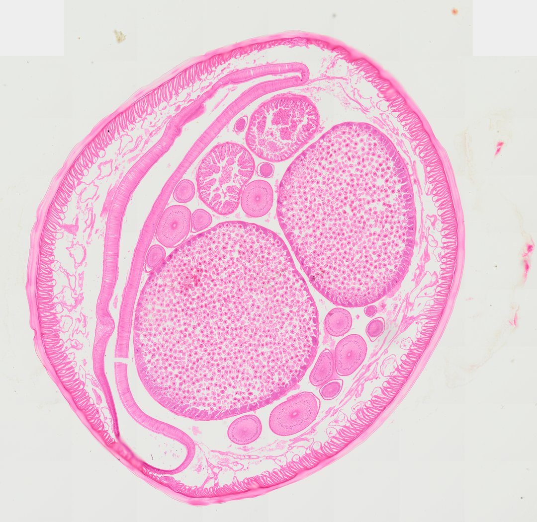
A cross section of a female Ascaris roundworm displaying classic pseudocoelomate anatomy. The specimen is bounded by a prominent, non-cellular thick cuticle overlying a thin hypodermis and tall, coelomyarian muscle cells projecting into the pseudocoel. The body cavity contains paired, large, thick-walled uterine branches filled with numerous fertilized eggs/ova, cross sections of coiled ovarian tubules containing developing oocytes, and a flattened intestine lined by simple columnar epithelial cells.
- Category
- Pathology & Parasitology
- Magnification
- 20x
- Stain
- H&E
- Tags
- Ascaris female cross section, Ascaris lumbricoides, Female roundworm, Nematoda, Pseudocoelomate, Uterus, Ovary, Oocytes, Fertilized ova, Cuticle, Coelomyarian muscle cells, Intestine, Columnar epithelium, Pseudocoelom, Helminthology, Cross section, Parasite histology, H&E
Explore this slide with deep zoom, save it to your library, and present it live to students on StudyScope. Create a teacher account or browse the full catalog.