Pine Needle (Cross Section)
Pinus
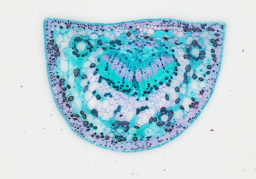
This cross section of a pine needle (Pinus leaf c.s.) demonstrates key anatomical adaptations characteristic of xerophytic gymnosperm foliage designed to minimize transpiration while maximizing photosynthetic efficiency. The outermost layer is enclosed by a thick cutinized cuticle covering a heavily sclerified epidermis and underlying hypodermis, which provides structural integrity and prevents water loss. Sunken stomata are embedded along the margin to regulate gas exchange under arid or freezing conditions. Internally, the mesophyll consists of distinctive plicate (infolded) parenchyma cells, increasing internal surface area for chloroplast distribution. Resin ducts surrounded by specialized epithelial secretory cells run longitudinally through the mesophyll. Enclosed by a prominent endodermis with a Casparian strip, the central vascular bundle contains transfusion tissue surrounding parallel xylem and phloem strands, facilitating water transport and nutrient trans-location.
- Category
- Botany & Plants
- Magnification
- 20x
- Stain
- Safranin and Fast Green
- Tags
- pine needle cross section, Pinus, gymnosperm anatomy, pine leaf c.s., plicate mesophyll, resin duct, sunken stomata, endodermis, transfusion tissue, xylem, phloem, hypodermis, thick cuticle, vascular bundle, xerophytic adaptation, conifer anatomy, plant histology, botanical microtechnique, fast green safranin stain, sclerified tissue, parenchyma, epidermis, plant tissue cross section
Explore this slide with deep zoom, save it to your library, and present it live to students on StudyScope. Create a teacher account or browse the full catalog.